
EKG/ Heart Monitoring
EKG or ECG records the electrical activity of the heart by attaching electrodes to the patient’s chest, arms and legs. It is usually performed in the doctor’s office or in emergency rooms. It records heart rhythm and gives a lot of useful information to the doctor. It may provide evidence of heart attacks or may even indicate previous silent heart attack. It may detect arrhythmias such as atrial fibrillation.
For further monitoring of heart rhythm, long-term heart monitors may be recommended. Continuous monitoring of the heart at home or at work can be achieved. 24 hour or 72 hour or weekly monitoring may be recommended.

Stress Test
To evaluate symptoms such as chest pain, shortness of breath, palpitations and fatigue, it is sometimes necessary to perform a stress test. Using electrodes placed on the body, this test monitors electrical activity and heart rhythm during exercise. It can help detect coronary artery disease, arrhythmias as well as exercise tolerance.
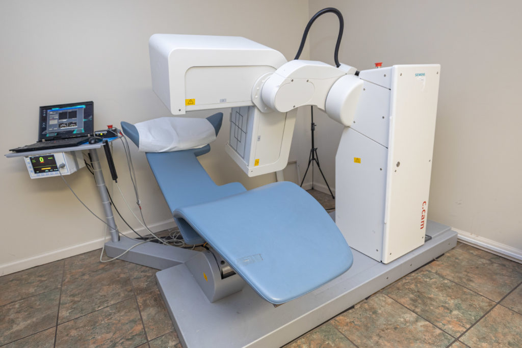
Nuclear Stress Test
When a simple stress test may not be adequate, a nuclear stress test may be ordered for evaluation. Scanning/imaging of the heart before and after exercise is performed to evaluate if there is any difference in blood flow to the heart muscle during exercise. Nuclear stress test gives additional information compared with simple stress test.
For patients who cannot exercise, chemical nuclear stress test is sometimes recommended. Scanning and imaging of the heart is done in a similar fashion.
The cardiologist usually decides which would be the most appropriate form of stress test depending on the patient and EKG.
Echo
An echocardiogram is performed to diagnose and manage certain heart conditions. It uses sound waves to create detailed images of the heart. It is a quick, painless and safe test. It can reveal a lot of useful information including damage to the heart muscle, valve problems, heart defects, heart size and heart function.

Carotid Doppler
Doppler testing uses high-frequency sound waves to perform a carotid ultrasound. This provides an image of blood flow in the carotid arteries in the neck. It is important because plaque buildup can narrow these arteries, located on either side of the neck and if severe can result in a stroke.
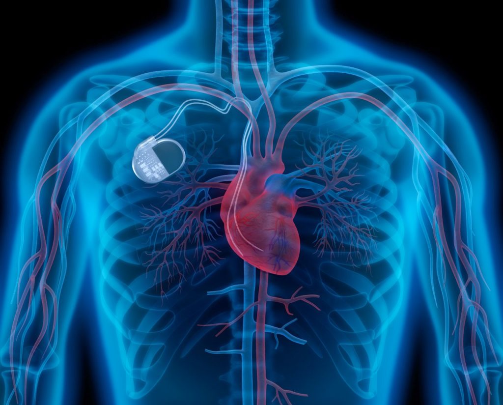
Pacemaker
A pacemaker is an electronic device that senses electrical activity of the heart/heart rhythm. It can also send electrical signals to the heart muscle to make it beat regularly. One of the common reasons for pacemaker insertion is if the heart is beating too slow. Pacemaker is usually inserted in the left upper or right upper chest below the skin. It consists of a metal case and electrical wires (leads) that are connected to the heart muscle. There are different types of pacemakers and cardiologists usually decide which type of pacemaker is needed depending on the patient’s condition.
Defibrillator
A defibrillator is a similar device like a pacemaker but usually larger in size. It can restore normal heartbeat/heart rhythm by sending an electric pulse or shock to the heart. Defibrillators have been useful in preventing sudden death in patients with severe heart damage or in patients known to have irregular heart rhythms such as ventricular tachycardia or ventricular fibrillation. Defibrillator is inserted in a similar fashion to a pacemaker.
Pacemaker Clinic
Pacemaker clinic is utilized for analysis, service and follow-up care of patient’s pacemakers/defibrillators as needed. Nowadays, some of the pacemakers / defibrillators are also followed up by remote monitoring
Heart Catheterization/Coronary Angiography
In heart catheterization (often called cardiac catheterization or cardiac cath) a very small, flexible, hollow tube (called a catheter) is placed in a blood vessel in the groin, arm or neck. Then it is advanced under x-ray guidance (fluoroscopy) into the aorta and towards the heart.
The catheter can be guided into the coronary arteries which supplies blood to the heart muscle. Contrast dye can be injected in the arteries to check for blockages/blood flow. This is called coronary angiography.
Angioplasty/Stent
If a significant blockage is found and which may be related to patient’s symptoms, then the cardiologist may recommend and may proceed with angioplasty where a balloon is utilized to open the blockage.
Stent, made out of wire mesh and mounted on a balloon, is usually placed after angioplasty in the coronary arteries. Stents help to prevent an artery narrowing again after angioplasty. Stents coated with medications are called drug-eluting stents and prevent further narrowing of blockages.
Angioplasty/stent procedures are also performed for blockages in other blood vessels besides coronary arteries.